- Use of microscope in dentistry: indications and peculiarities
- When is a dental microscope used?
- Dental diagnostics under the microscope
- Determining the location of dental canals with a microscope
- Treatment of tooth canals under the microscope
- Tooth canal filling
- Benefits of dental treatment under the microscope
- Frequently asked questions about dental treatment and root canals under a microscope
- Specialists
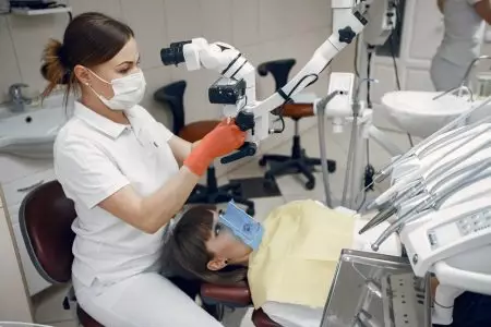
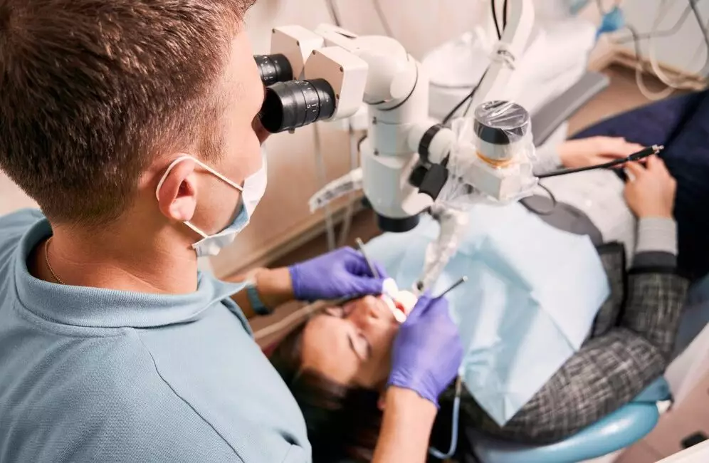
Treatment of teeth and dental canals under a microscope is a specialised method in which the dentist uses a dental microscope to magnify and illuminate the working area during the procedure. The method is actively used in the clinic “Dent-House” in Odessa. Our doctors carry out accurate and effective manipulations in any cases – make an appointment right now!
Use of microscope in dentistry: indications and peculiarities
Using a microscope in dentistry is like using a very powerful magnifying glass to help the doctor look at teeth and dental canals in more detail.
When a dentist uses a microscope, they can see even the smallest problems on your teeth. This means that the treatment will be better and more successful, which in turn will help keep your smile healthy and beautiful for a long time. The cost of treatment will be a little higher, but the price is well worth the quality of the procedure – positive patient reviews confirm this.
When is a dental microscope used?

Dental microscope in Odessa is used in therapeutic, endodontic, orthopaedic and aesthetic dentistry. With its help, the dentist can:
- detect early caries, microcracks and chips that are not visible even on X-rays;
- to clean and treat dental problems;
- to treat the roots of the tooth;
- to seal the fissures;
- to restore teeth;
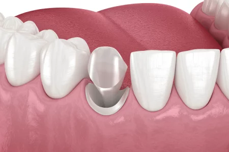
- to treat teeth for crowns and veneers;
- remove old fillings, cysts and pins;
- to put in implants.
The application of the dental microscope is quite wide. You can find out how much treatment costs at Dent-House Clinic by opening the “Prices” tab.
Dental diagnostics under the microscope
Under a microscope, the dentist can detect the initial stages of decay, cracks, scuffs and other damage to teeth that can lead to more serious problems and diseases in the future. Microscopic dental diagnostics can be used to evaluate root canals and plan implants.
This diagnostic method helps the dentist develop a more effective and personalised treatment plan for each patient, and in this way ensures that teeth are preserved for years to come.
Determining the location of dental canals with a microscope
Dental canals are narrow cavities inside the tooth that are located in the root of the tooth. The tooth has one or more canals filled with pulp – connective tissue, nerves and blood vessels. The dental canals serve to transport nutrients and oxygen to the living tooth tissue, and to remove metabolic waste and decay products from the tissue.
When a tooth becomes decayed, traumatised or infected, the tooth canals can also become damaged. In such cases, canal treatment may be required, which includes removing the damaged pulp and cleaning the canals of bacteria and other harmful substances. The procedure may also be required due to trauma, periodontal disease, or cracks in the tooth.
Under the microscope, the doctor can see the structure of the tooth as well as the depth of the canal thanks to the 40-fold magnification of the image. This is especially important in case of complex or non-standard anatomical features of the teeth.
Treatment of tooth canals under the microscope
The procedure of endodontic treatment of a tooth under a microscope in Odessa usually goes like this:
- The patient is given a local anaesthetic to make them comfortable during the procedure.
- The tooth is isolated from saliva using a cofferdam.
- The dentist removes the affected tissue and carious formations with a dental drill. The canals of the tooth are then accessed.
- The doctor carefully examines the tooth canals with high magnification to detect any abnormalities or problems.
- The dentist removes the pulp and cleans the canals of the tooth using special instruments.
- The canals are dilated and flushed with disinfectant solutions.
The procedure of treating tooth canals under a microscope requires special attention and skills on the part of the dentist.
Tooth canal filling

Tooth canal filling is a procedure that is performed after treatment.
There are several types of canal filling materials, but gutta-percha is commonly used. A post is also placed in the smooth, wide canals and the space around it is filled with gutta-percha.
- After selecting a suitable composition, the dentist will fill the roots of the tooth. The material is inserted firmly into the root canals using special thin instruments to fill them completely.
- The dentist then makes sure that the material is evenly and correctly distributed. This is usually done by taking X-rays. A temporary or permanent filling is then placed on the tooth.
Tooth canal filling helps prevent bacteria and infection from re-entering the tooth and preserving its integrity.
You can see how much it costs to have a root canal filling on our Dent-House website. Our receptionist will also tell you the cost when you make an appointment. However, please note that the price is approximate: depending on your individual case, the doctor will be able to work out the full price only at your appointment during an examination before treatment.
Benefits of dental treatment under the microscope
Endodontic treatment of teeth under a microscope has several advantages:
- With a microscope, the dentist can take a closer look at the teeth and their structure.
- Thanks to its high magnification and image quality, the microscope allows the dentist to detect even the smallest defects and problems on the surface of teeth and in root canals.
- The use of a microscope improves the quality of dental treatment because the dentist can clean and treat the teeth more thoroughly.
- The dentist works with greater precision and control. This reduces the risk of damage to surrounding tissues and complications during the procedure.
- Thanks to the microscope, high-quality tooth restorations can be carried out.
- It is possible to correct a problem even if the previous dentist made a mistake in treatment.
In general, dental treatment under a microscope provides greater accuracy, efficiency, and safety in various procedures. Customer feedback suggests that the treatment is not only painless and comfortable, but also effective.
Trust root canal treatment and other procedures, such as whitening, to the experts at Dent-House Family Dentistry in Odessa. Make an appointment to receive quality treatment without pain and worry.
Frequently asked questions about dental treatment and root canals under a microscope
🦷 How to prepare for treatment under a microscope?
😁 How long does root canal treatment under a microscope take?
🦷 Is it possible to treat teeth under a microscope if there is severe pain?
😁 Is a follow-up appointment necessary after root canal treatment under a microscope?
Cost of services
Specialists

Semenyuk Ekaterina Sergeevna
Dentist

Kirilyuk Yulianna Ivanovna
Pediatric dentist

Furduy Ekaterina Sergeevna
Pediatric dentist


